#ICYMI: Study links cannabis use to lower risk of cognitive decline, dementia-related diseases — via @healthing_ca#Dementia #Study #Cannabis https://t.co/R9WwSFVqtq
— The Vancouver Sun (@VancouverSun) April 21, 2024
Neuroscience
https://www.sciencealert.com/keto-diet-may-slow-down-alzheimers-mouse-study-reveals10
April 2024
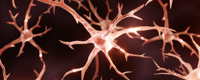 (Artur Plawgo/Getty Images)
We know that a host of factors play into Alzheimer’s risk, including the makeup of our gut bacteria. So, it makes sense that our diets might have some influence.
A new study found a chemical elevated by ‘ketogenic’ diets – which are low in carbs and higher in proteins and fats – can delay the early stages of Alzheimer’s-related memory loss in mice. That memory loss is comparable to the mild cognitive impairment seen in people before Alzheimer’s takes hold.
Key to the new discovery by a team led by the University of California, Davis (UCD), is the molecule beta-hydroxybutyrate (BHB), which the ketogenic diet increases levels of. Here, the researchers found BHB was particularly abundant in biological pathways associated with memory and brain plasticity.
“The data supports the idea that the ketogenic diet in general, and BHB specifically, delays mild cognitive impairment and it may delay full blown Alzheimer’s disease,” says biochemist Gino Cortopassi, from UCD.
The ketogenic diet has long been linked to weight loss and other health benefits. Indeed, some of the same researchers had already found in mice that it’s associated with longer lives and staying healthier for longer.
The diet shifts the body’s metabolism, so instead of using glucose for energy, it burns dietary and stored fat, producing ketones for fuel instead of glucose. Without elevated glucose levels, insulin levels no longer peak and crash after meals, reducing the ensuing hunger cravings.
In this study, the extra BHB produced by the keto diet was shown to have a positive effect on the brain, effectively keeping its gears turning – gears that can become slower over time and into old age.
“We observed amazing abilities of BHB to improve the function of synapses, small structures that connect all nerve cells in the brain,” says Izumi Maezawa, a pathologist at UCD.
“When nerve cells are better connected, the memory problems in mild cognitive impairment are improved.”
We’ve already seen links between ketogenic diets and neurological conditions like epilepsy – so there’s some kind of interplay going on here. However, high levels of ketones come with their own health risks, and scientists aren’t sure just how good keto diets are for us in general, especially over the long term.
While we might not see doctors asking patients to drastically change how they eat, the findings should help us better understand how to block or slow down the changes in the brain that lead to Alzheimer’s.
Another important point from the study – the keto diet produced more BHB in female mice and seemed to benefit them more. The role of gender in Alzheimer’s risk is another aspect of the disease scientists are working to explain.
“If these results translated to humans, that could be interesting since females, especially those bearing the ApoE4 gene variant, are at significantly higher risk for Alzheimer’s,” says Cortopassi.
The research has been published in Communications Biology.
Data from almost 100 million people compares the expected and observed incidents of side effects
https://www.rt.com/news/592695-covid-vaccine-effects-study
Feb 19, 2024

File photo © Getty Images/Toshe_O
A big data study of 99 million people across eight countries showed greater than expected incidence of side effects from various Covid-19 vaccines, the Global Vaccine Data Network (GVDN) said on Monday.
The study, originally published in the medical journal Vaccine on February 12, looked at 13 neurological, blood, and heart-related conditions, called “adverse events of special interest.” Researchers looked at 99,068,901 vaccinated individuals from ten sites in eight countries.
“The size of the population in this study increased the possibility of identifying rare potential vaccine safety signals,” said Kristyna Faksova, the lead author of the study from the Statens Serum Institut in Copenhagen, Denmark.
According to the GVDN, the study observed a greater incidence of myocarditis (inflammation of the heart muscle) and pericarditis (inflammation of the heart sac) than expected among those who took the Pfizer/BioNTech (BNT162b2) and Moderna (mRNA-1273) shots.
Moderna’s vaccine also had a higher rate of acute disseminated encephalomyelitis (ADEM, inflammation and swelling in the brain and spinal cord), with seven observed events compared to two expected within 42 days of the first shot.
Russia changes mandatory Covid-19 vaccination rules
Safety signals for myocarditis were “consistently identified” following the first three doses of either mRNA shot, with the highest ratio after the second dose. Signals for pericarditis also appeared following the first and fourth doses of mRNA-1273 and were also observed after a third dose of the Oxford/Astra Zeneca (ChAdOx1) viral vector vaccine.
Recipients of ChAdOx1 had 190 observed events of Guillain-Barré Syndrome (GBS) compared to the expected 76, and 69 observed instances of cerebral venous sinus thrombosis (CVST, a type of blood clot in the brain) compared to the expected 21, the study has shown.
The GVDN has made the results available to the public on its interactive data dashboards, along with a warning not to read too much into correlations and that the vaccines are both safe and effective.
“By making the data dashboards publicly available, we are able to support greater transparency, and stronger communications to the health sector and public,” GVDN co-director Dr. Helen Petousis-Harris said.
The study was part of the GVDN’s Global COVID Vaccine Safety Project and was funded entirely by a $10 million grant from the US Centers for Disease Control and Prevention (CDC).
A study published in the Nutrition Journal found an association between regular consumption of sugary beverages and dementia risk. The study found that free sugars in beverages can increase dementia risk by upwards of 39 percent.
https://herbs.news/2024-01-12-research-shows-lions-mane-mushroom-combats-dementia.html
01/12/2024
Currently, there are more than 55 million people who suffer from dementia worldwide, and nearly 10 million new cases of dementia are diagnosed each year. Cognitive decline has become such a pervasive issue in modern society; it has become normalized across the political spectrum. Some of today’s government officials show serious cognitive decline, and even the de facto President of the United States routinely stumbles around in a stupor, taking cues from handlers and mumbling incoherently at times.
Cognitive decline is a serious health issue worldwide, but in many cases, there are ways to reverse the damage, prevent the death of neurons and regenerate neuronal pathways. Lion’s mane mushroom is an important medicinal food that can promote the biosynthesis of nerve growth factor and effectively combat dementia.
Lion’s mane mushroom promotes the biosynthesis of nerve growth factor
A study published in Mycology finds that Lion’s mane mushroom (Hericium erinaceus) synthesizes two very important compounds for nerve growth – Hericenones and erinacines. These compounds are derived from the fruiting body and mycelium of the mushroom. Both compounds promote the biosynthesis of nerve growth factor (NGF) and therefore have value in the prevention and treatment of dementia.
Scientists have isolated two erinacine derivatives and two erinacine diterpenoids (Cyatha-3 and 12-diene with isomer) that promote NGF. Scientists have also demonstrated NGF-stimulating activity from three other compounds in Lion’s mane – Hericenones C, D and E. One of the compounds, 3-Hydroxyhericenone F, showed protective activity against endoplasmic reticulum stress-dependent Neuro2a cell death.
Two other species of the mushroom contained several compounds that promote nerve growth factor. Sarcodon scabrosus (A-F) and Sarcodon cyrneus (A-I, P, Q, J, R, K) all show promise for prevention and treatment of cognitive decline.
Interestingly, both the Hericenones and erinacines are low-molecular weight compounds that cross the blood-brain barrier with ease. The lion’s mane mushroom was designed at the molecular level to positively affect the brain and heal the nervous system, promoting peripheral nerve regeneration and advancing learning abilities into old age.
High blood sugar levels harden arteries, increasing risk of dementia
While there are ways to reverse cognitive decline through medicinal foods, the prevention of dementia should always be approached through a holistic perspective. When treating dementia, it’s equally important to eliminate the chemicals that are promoting cognitive decline. High blood sugar levels are known to harden the arteries, increasing the risk of blockages in the brain. Obstruction of blood flow to the brain can inhibit blood supply to the nerve cells, resulting in impaired brain function.
A study published in the Nutrition Journal found an association between regular consumption of sugary beverages and dementia risk. The study found that free sugars in beverages can increase dementia risk by upwards of 39 percent. The study included a dietary analysis from 186,622 participants from the UK Biobank cohort. The analysis spanned 206 types of food and 32 types of beverages consumed over the course of 10.6 years. The analysis found a correlation between fructose, glucose and sucrose (table sugar) and dementia risk. The free sugars in soda, fruit drinks and milk-based drinks were strongly related to dementia risk, while the sugars in tea and coffee showed minimal risk.
Herbal teas – including but not limited to: green tea, chamomile, lavender and lemon balm – are all wonderful alternatives to sugar-laden drinks. These beverages, when sweetened with plant-based stevia extract, also provide the body with antioxidants, polyphenols and theaflavins that fight free radicals and therefore protect the brain.
Dementia doesn’t have to plague the population and dumb down the people who are running our government and institutions. Advanced learning can continue into old age. Herbal teas can replace sugary beverages in the diet, thus protecting the brain. Medicinal foods like lion’s mane mushroom can heal damaged neurons while promoting new neuron growth.
Sources include:
FungalBiotech.org [PDF]
When it goes online next year, it will be capable of 228 trillion synaptic operations per second, which rivals the estimated rate of operations in the human brain.
Ellie Zolfagharifard
Business Insider
Fri, 15 Dec 2023

Our brains are remarkably energy efficient.
Using just 20 watts of power, the human brain is capable of processing the equivalent of an exaflop — or a billion-billion mathematical operations per second.
Now, researchers in Australia are building what will be the world’s first supercomputer that can simulate networks at this scale.
The supercomputer, known as DeepSouth, is being developed by Western Sydney University.
When it goes online next year, it will be capable of 228 trillion synaptic operations per second, which rivals the estimated rate of operations in the human brain.
The hope is to better understand how brains can use such little power to process huge amounts of information.
If researchers can work this out, they could someday create a cyborg brain vastly more powerful than our own. The work could also revolutionize our understanding of how our brains work.
“Progress in our understanding of how brains compute using neurons is hampered by our inability to simulate brain-like networks at scale,” said André van Schaik, a director at Western Sydney University’s International Centre for Neuromorphic Systems.
“Simulating spiking neural networks on standard computers using Graphics Processing Units and multicore Central Processing Units is just too slow and power intensive,” he added. “Our system will change that.”
Ralph Etienne-Cummings at Johns Hopkins University, Baltimore, who is not involved in the work, told New Scientist that DeepSouth will be a game changer for the study of neuroscience.
“If you are trying to understand the brain this will be the hardware to do it on,” he said.
Etienne-Cummings said that there will be two main types of researchers who will be interested in the technology — those studying neuroscience, and those who want to prototype new engineering solutions in the AI space.
DeepSouth is just one of many research projects aiming to create a machine that will rival the human brain.
Other researchers are trying to tackle the same problem by creating “biological computers” powered by actual brain cells.
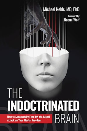
My Foreword to Bombshell New Book, ‘The Indoctrinated Brain’
https://www.globalresearch.ca/has-neuroscience-found-key-what-caused-zombie-nation/5842223
By Dr. Naomi Wolf and Dr. Michael Nehls
Global Research, December 05, 2023

***
Please enjoy – to the extent you can – my Foreword to ‘The Indoctrinated Brain’, by Dr Michael Nehls. The book, which explains the neuroscience of propaganda, will chill your soul but may explain a great deal.
‘The fact that the brain is plastic—modifiable—has become much better understood by the public in the past few decades.
General readers understand by now that the human brain can be altered; and that experiences can modify its reactions and processes. We understand now for example that PTSD leaves lasting changes in brain functioning. It’s been established that motherhood changes the brain and that bonding itself is a chemical process modified by the brain.
We also understand, as general readers, that propaganda is real.
Some of us have studied propaganda in the past. We have a working knowledge of Joseph Goebbels, and of the artistry and craft that underlay his manufacturing of National Socialist consent. The work of Edward Bernays, one of the earliest practitioners of what became the field of public relations, has been widely read in English. Decades-old bestsellers such as Subliminal Seduction by Wilson Bryan Key exposed the fact that advertisers use every tool at their disposal to alter our reactions to their products—down to the level of the subconscious mind.
Modern general audiences also understand that governments use “messaging”—and often, heavy-handed propaganda—to lead us to take actions that can be against our interests or our better conscious judgments; to create prejudices and divisions that may not otherwise exist; to heighten fears and to trigger a sense of vulnerability in us, so that we can be better manipulated and guided to goals that are not our own.
But Dr. Michael Nehls’s thesis in this book is revolutionary because it brings together all of these fields of inquiry and proposes a set of questions so radical that they make the mysteries of the past three years fall into place. This is the indispensable book. In The Indoctrinated Brain, Dr. Nehls brings these areas of study together in a way that has never been done before. By applying neuroscience to the otherwise bizarre events of the recent past, he explains what has happened to humanity.
Many of us have noted that our loved ones and colleagues have changed. Post- mRNA injection rollout, we notice that people who were highly educated critical thinkers have become unable to think outside of two simple binaries. We watch in astonishment as formerly sophisticated loved ones and friends regurgitate talking points with no self-awareness. We wonder why there is a sense of something inchoately missing when we sit with a vaccinated or COVID-fearful friend. We cannot fathom what has caused this sea change.
Dr. Nehls’s hypothesis can explain it.
“The Indoctrinated Brain introduces a largely unknown, powerful neurobiological mechanism whose externally induced dysfunction underlies these catastrophic developments,” as the publisher notes.
Dr. Nehls argues that the spike protein, along with other COVID measures, represents an intentional attack on the human hippocampus—where autobiographical memory and individuality itself originate—and that “fear porn” keeps us from holding on to the autobiographical memories that encompass our former selves. As a result, humans have become deindividualized, more suggestible, more forgetful, more compliant, and less able to engage in critical thinking and creative reasoning. This argument utterly accords with what many of us are seeing, to our horror, every day. Dr. Nehls’s The Indoctrinated Brain is an indispensable book because it applies neuroscience to politics and especially to the politics of fascism. The need for that has existed for as long as modern fascism has existed.
Neuroscience should be applied to politics and to social change, but it is rare indeed when those fields of analysis meet. By bringing these fields of knowledge together and mapping neurological science against propaganda, and vice versa, Dr. Nehls brings vast new insights to the reader that would not have been attainable previously.
After you read The Indoctrinated Brain, you will think: Of course. Of course, the propaganda of the past few years must have been predicated upon intensive study of the brain and its reactions. Of course, the hundreds of millions of dollars that were recently spent and are currently being spent by the US and other governments on behavioral science and behavior modification would result in insights that would be applied by the US and other governments to making populations more tractable, less able to reason, less creative and more compliant.
Why else would they so heavily have invested in such studies? Of course, the constant messaging, especially about fear, over the past three years, would have an effect that is not just about public health or perhaps not at all about public health—but that it is rather about making humans in free societies more tractable—with public health as the excuse, the proxy, for this deployment of life-altering and consciousness-altering fear. It is not the fear porn about the specific scary thing that matters, Dr. Nehls persuasively argues here: the fear itself is deliverable. The fear itself changes and indeed damages the brain.
I’ve long been interested in the psychiatric effects and, as I guessed, intentionalities behind “lockdowns” and “pandemic” messaging. But I did not have the neuroscientific background to understand exactly what was being done to people via “lockdowns” and the “fear porn” of the pandemic years related to the virus—to other human beings.
Through my study of the psychiatric effects of torture and isolation, that I took on for a book about closing democracies, I realized that isolation causes profound and sometimes permanent changes in the brain. I knew intuitively in the post-9/11, “Global War on Terror” years, that constant fear would wear down faculties needed for critical thinking. And I applied those insights to the isolation and fear messaging of 2020–22. But I did not have the complete picture.
This book provides it. It is the “aha” hypothetical for our time.
The Indoctrinated Brain provides the missing practical knowledge of neuroscience, that explains why isolating people creates a more befuddled, more easily manipulated population. It explains exactly why a message that closeness with other human beings can kill you, or you can kill others (especially your grandma) through physical closeness, might rewire the human brain to create the vulnerability to delusion and bad science and cultlike thinking, that many of us observed in formerly critically thinking loved ones and friends, post-2020. It even raises the question of whether the spike protein contributes to brain fog and to the erasure of a sense of an autonomous, resilient, individuated, and questing self.
If Dr. Nehls is right, his theory here will be as important as Dr. Sigmund Freud’s discovery of the subconscious, if not more so. If he is right, his theory explains why governments around the world mandated “lockdown” measures and mRNA injections, which would not ultimately then be about public health but about creating manipulable, passive citizens.
If Dr. Nehls is right, it explains so many baffling features of the past three years—notably the fact that formerly thoughtful, highly individuated leaders of institutions, down to rank-and-file citizens, followed cultlike dicta without a mur- mur, and pursued nonsensical goals such as isolation, masking, and submission to vaccine mandates, without protest. Dr. Nehls’s thesis would explain the bizarre experience many of us are having of watching our formerly analytical loved ones, find themselves unable to keep two thoughts in their heads at the same time, unable to engage in calm debate without exploding emotionally, unable to maintain contact and connection with people with whom they disagree.
As I write, another global crisis is being spun up, this one in the Middle East. Within a day, highly educated and formerly skeptical loved ones of mine are repeating glaring legacy media talking points without any self-consciousness. It’s upsetting not to know why they would change in this way—and it is even more upsetting, though incredibly enlightening, to read Dr. Nehls’s argument and realize what the cause may be of their submissiveness to propaganda narratives. It makes it both easier and harder to contend with loved ones, friends, and colleagues who have been intellectually blunted in this way, to understand Dr. Nehls’s point of view and realize that this sad change in cognition might be simply physical—the spike protein—and neuropsychiatric: the repetition of fear messages and their impact on the brain.
In my social media feed today—on a day when the news has brought images of endless atrocities to our media streams, and when we are being told that this Friday will be a “Day of Jihad” with plenty of stabbings—someone wrote, “Protect your amygdala.” That meant, do not expose yourself to endless scenes of rape, murder, beheadings, atrocities, and horrors.
Dr. Nehls’s book is ultimately a hopeful one, since if we understand the damage to our brains from both spike proteins and fear pornography, we can find ways to prospect ourselves and our conscious minds. I appreciate the practical suggestions Dr. Nehls gives us to do just that.
It is scary that we are living in a time in which there is, as Dr. Nehls so powerfully points out, a war on our brains. But it must be less scary to understand what is being done to us, with Dr. Nehls’s help, so we can protect and strengthen our autobiographical memory and critical thinking, and so we can survive this onslaught with the full range of our intelligence—and our humanity—intact.‘
The Indoctrinated Brain
How to Successfully Fend Off the Global Attack on Your Mental Freedom
By Michael Nehls, Naomi Wolf
Global War on the Human Brain
Throughout the world, mental capacity is declining, especially among young people, while depression rates are rising dramatically. Meanwhile, one in forty men and women suffers from Alzheimer’s, and the age of onset is falling rapidly. But the causes are not being eliminated, quite the opposite. Can this just be coincidence?
The Indoctrinated Brain introduces a largely unknown, powerful neurobiological mechanism whose externally induced dysfunction underlies these catastrophic developments.
Michael Nehls, medical doctor and internationally renowned molecular geneticist, lays out a shattering chain of circumstantial evidence indicating that behind these numerous negative influences lies a targeted, masterfully executed attack on our individuality. He points out how the raging wars against viruses, about climate change, or over national borders are—more likely intended than not—fundamentally providing the platform for such an offensive against the human brain that is steadily changing our being and is aimed at depriving us of our ability to think for ourselves.
But it is not too late. By exposing these brain-damaging processes and describing countermeasures that anyone can take, Nehls brings light and hope to this fateful chapter in human history. Nothing less will be decided than the question of whether our species can retain its humanity and its creative power or whether it will lose them irretrievably.
- 288 Pages
- December 12, 2023
- ISBN: 9781510778368
- Imprint: Skyhorse Publishing
- Trim Size: 6in x 9in x 0in
The original source of this article is Outspoken with Dr Naomi Wolf
Copyright © Dr. Naomi Wolf and Dr. Michael Nehls, Outspoken with Dr Naomi Wolf, 2023
In encouraging news for people recovering from alcohol use disorder, new research demonstrates how quickly the brain can repair its structure once drinking ceases.
09 November 2023
 (PhonlamaiPhoto’s Images/Canva Pro)
In encouraging news for people recovering from alcohol use disorder, new research demonstrates how quickly the brain can repair its structure once drinking ceases.
People with alcohol use disorder (AUD) tend to have thinning in regions of their cortex; the wrinkled outer layer to the brain critical to so many higher-order cognitive functions. The US study found those who quit drinking gain cortical thickness over time, faster in the first month and continuing over 7.3 months, at which point thickness is comparable to those without AUD.
Previous research has shown that some regions may recover when someone stops drinking, but it was unclear how much or how quickly recovery occurs.
“The few longitudinal studies investigating cortical thickness changes during abstinence are limited to the first month of sobriety,” writes the team, led by psychiatrist and behavioral scientist Timothy Durazzo from Stanford University.
“However, the extent of regional cortical thickness recovery over an extended period of abstinence (e.g., greater than 6 months) is unknown.”
An estimated 16 million people in the US suffer from AUD. It’s a major public health issue, and understanding this complex disorder is important for treatment, prevention, and reducing stigma.
Alterations to brain structure and function during chronic alcohol use can make it tough for people to stop drinking, despite their best intentions. For instance, the prefrontal cortex – an area involved in planning and decision-making – may become less active, making it harder for people with AUD to make healthy decisions.
Durazzo and colleagues also examined how some health conditions, smoking history, psychiatric conditions, and substance use disorders affect longer-term cortical thickness changes in people recovering from AUD.
Altogether, 88 people with AUD participated in the study, undergoing brain scans at approximately 1 week, 1 month, and 7.3 months of abstinence. Some participants joined at the 1-month mark, meaning 23 individuals didn’t have scans taken at 1 week, and only 40 of the total 88 continued to abstain from alcohol for the full period.
They also looked at 45 people who had never had AUD, measuring their cortical thickness at baseline and again about 9 months later to confirm the areas that were measured stayed the same.
A type of magnetic resonance imaging ( MRI) that’s particularly useful for getting clear pictures of the body’s internal structure was used to observe the participants’ brains. The researchers recorded cortical thickness for 34 regions, averaging the measurement across the brain’s left and right hemispheres.
Recovery of thickness in those with AUD after 7.3 months without alcohol was quite widespread. It was enough to be statistically significant in 25 of the 34 regions, and 24 of these were considered statistically equivalent in thickness to controls.
All 34 cortical regions that Durazzo and his team looked at saw a faster rate of thickness change in AUD participants from 1 week to 1 month after quitting than from 1 month to 7.3 months.
Cortical thickening happened more slowly in some parts of the brains of people with AUD who also had high blood pressure or high cholesterol. The same was true for people with AUD who were current smokers.
No significant relationships were found between cortical thickness changes and current substance abuse (including drugs other than alcohol), psychiatric disorders, or past cigarette smoking. So quitting smoking might also contribute to the recovery of cortical thickness.
These results provide encouragement and a new understanding of brain recovery after quitting alcohol, though due to the small sample size and lack of diversity, they may not be generalizable. Also, it’s important to note these findings don’t indicate whether the changes had any effect on brain function.
“Larger longitudinal studies are required to examine the neurocognitive and psychosocial correlates of cortical thickness recovery during sustained abstinence in AUD,” the team writes.
The authors also point out that variables they didn’t account for, such as genetics, physical activity, and people’s liver and lung health, could have affected their findings.
“This data provides clinically relevant information on the beneficial effects of sustained sobriety on human brain morphology,” the authors conclude, “and reinforces the adaptive effects of abstinence-based recovery in AUD.”
The study has been published in Alcohol.
The team ran a trial involving 72 adults aged over 60 across the course of 12 weeks. The participants were split randomly and unknowingly into two groups: the first group took a wasabi tablet once a day, while the second group took a placebo tablet.
At the end of the experiment, those who had ingested wasabi tablets showed significantly better performance in their episodic memory (recalling events from the past) and working memory (holding information temporarily) based on a series of cognitive tests.
https://www.sciencealert.com/wasabi-boosts-cognitive-ability-in-older-people-study-shows
03 November 2023
 (Aksana Ban/Moment/Getty Images)
A new study shows that the traditional Japanese spice wasabi can improve some types of cognitive function in those aged over 60, offering a straightforward and rather tasty way of looking after our brains as we get older.
Key to the association is the wasabi ingredient 6 methylsulfinyl hexyl isothiocyanate (6-MSITC), a bioactive compound that has previously been linked to antioxidant and anti-inflammatory effects that slow down damage to cells and protect them.
While a small number of studies have shown 6-MSITC having a beneficial effect on cognition, this hasn’t yet been tested with older adults – the people who are most at risk of cognitive decline and related problems like dementia.
“Previous studies suggested that antioxidants and anti-inflammatories have an important role in cognitive health in older adults,” write the researchers in their published paper. “Therefore, 6-MSITC is expected to have a positive effect on cognitive performances in older adults.”
The team ran a trial involving 72 adults aged over 60 across the course of 12 weeks. The participants were split randomly and unknowingly into two groups: the first group took a wasabi tablet once a day, while the second group took a placebo tablet.
At the end of the experiment, those who had ingested wasabi tablets showed significantly better performance in their episodic memory (recalling events from the past) and working memory (holding information temporarily) based on a series of cognitive tests.
However, no significant differences were found in other areas of cognitive performance, including reasoning, attention, and processing speed. The team thinks the wasabi, and 6-MSITC, might be particularly affecting the hippocampus part of the brain, which is particularly important for memory function.
“These findings suggest that the 12 weeks’ 6-MSITC intake selectively enhances working and episodic memory functions in healthy older adults,” write the researchers.
The team now wants to look in more detail at what might be happening on the biological and molecular level. In this particular study, no antioxidant or anti-inflammatory biomarkers were measured, so it’s only possible to hypothesize about the effect the wasabi is having and why it’s having that effect.
Even with those limitations though, the study shows a clear link between the 6-MSITC-containing wasabi and better memory function. When it comes to maintaining healthy brains into old age, picking foods that are particularly good for us and avoiding those that aren’t can be a simple, relatively easy-to-follow strategy.
“Older adults with lower cognitive performances feel difficulties in daily behaviors such as shopping, banking, and cooking,” write the researchers.
“Therefore, it is important to improve cognitive functions in older adults.”
The research has been published in Nutrients.
See here:
Summary: A new study reveals that culture and geography significantly influence sleep patterns. Using data from Nokia smartwatches, the researchers analyzed 52 million logs over four years from 30,082 individuals in 11 countries.
The study highlighted that sleep-time varied per country, with high-GDP countries recording delayed bedtimes. Individual efforts like daily exercise can counterbalance cultural influences, with effects varying by country.
Key Facts:
- The study used extensive data from Nokia smartwatches, providing a detailed analysis of sleep patterns across 11 countries.
- Cultural and geographical factors significantly influence sleep patterns, with high-GDP countries and collectivist cultures recording delayed bedtimes.
- Daily exercise can counterbalance these cultural influences and improve sleep quality, but its effectiveness varies by country.
Source: KAIST
Sleep has a huge impact on health, well-being and productivity, but how long and how well people sleep these days has not been accurately reported.
Previous research on how much and how well we sleep has mostly relied on self-reports or was confined within the data from the unnatural environments of the sleep laboratories.
So, the questions remained: Is the amount and quality of sleep purely a personal choice? Could they be independent from social factors such as culture and geography?

A new study led by researchers at Korea Advanced Institute of Science and Technology (KAIST) and Nokia Bell Labs in the United Kingdom investigated the cultural and individual factors that influence sleep.
In contrast to previous studies that relied on surveys or controlled experiments at labs, the team used commercially available smartwatches for extensive data collection, analyzing 52 million logs collected over a four-year period from 30,082 individuals in 11 countries.
These people wore Nokia smartwatches, which allowed the team to investigate country-specific sleep patterns based on the digital logs from the devices.
< Figure comparing survey and smartwatch logs on average sleep-time, wake-time, and sleep durations. Digital logs consistently recorded delayed hours of wake- and sleep-time, resulting in shorter sleep durations. >
Digital logs collected from the smartwatches revealed discrepancies in wake-up times and sleep-times, sometimes by tens of minutes to an hour, from the data previously collected from self-report assessments.
The average sleep-time overall was calculated to be around midnight, and the average wake-up time was 7:42 AM. The team discovered, however, that individuals’ sleep is heavily linked to their geographical location and cultural factors.
While wake-up times were similar, sleep-time varied by country. Individuals in higher GDP countries had more records of delayed bedtime. Those in collectivist culture, compared to individualist culture, also showed more records of delayed bedtime.
Among the studied countries, Japan had the shortest total sleep duration, averaging a duration of under 7 hours, while Finland had the longest, averaging 8 hours.
Researchers calculated essential sleep metrics used in clinical studies, such as sleep efficiency, sleep duration, and overslept hours on weekends, to analyze the extensive sleep patterns. Using Principal Component Analysis (PCA), they further condensed these metrics into two major sleep dimensions representing sleep quality and quantity.
A cross-country comparison revealed that societal factors account for 55% of the variation in sleep quality and 63% of the variation in sleep quantity.
Countries with a higher individualism index (IDV), which placed greater emphasis on individual achievements and relationships, had significantly longer sleep durations, which could be attributed to such societies having a norm of going to bed early.
Spain and Japan, on the other hand, had the bedtime scheduled at the latest hours despite having the highest collectivism scores (low IDV).
The study also discovered a moderate relationship between a higher uncertainty avoidance index (UAI), which measures implementation of general laws and regulation in daily lives of regular citizens, and better sleep quality.
Researchers also investigated how physical activity can affect sleep quantity and quality to see if individuals can counterbalance cultural influences through personal interventions.
They discovered that increasing daily activity can improve sleep quality in terms of shortened time needed in falling asleep and waking up. Individuals who exercise more, however, did not sleep longer.
The effect of exercise differed by country, with more pronounced effects observed in some countries, such as the United States and Finland. Interestingly, in Japan, no obvious effect of exercise could be observed.
These findings suggest that the relationship between daily activity and sleep may differ by country and that different exercise regimens may be more effective in different cultures.
This research published on the Scientific Reports by the international journal, Nature, sheds light on the influence of social factors on sleep.
One of the co-authors, Daniele Quercia, commented: “Excessive work schedules, long working hours, and late bedtime in high-income countries and social engagement due to high collectivism may cause bedtimes to be delayed.”
Commenting on the research, the first author Shaun Sungkyu Park said, “While it is intriguing to see that a society can play a role in determining the quantity and quality of an individual’s sleep with large-scale data, the significance of this study is that it quantitatively shows that even within the same culture (country), individual efforts such as daily exercise can have a positive impact on sleep quantity and quality.”
“Sleep not only has a great impact on one’s well-being but it is also known to be associated with health issues such as obesity and dementia,” said the lead author, Meeyoung Cha.
“In order to ensure adequate sleep and improve sleep quality in an aging society, not only individual efforts but also a social support must be provided to work together,” she said.
“The research team will contribute to the development of the high-tech sleep industry by making a code that easily calculates the sleep indicators developed in this study available free of charge, as well as providing the benchmark data for various types of sleep research to follow.
About this sleep research news
Author: Yoonju Hong
Source: KAIST
Contact: Yoonju Hong – KAIST
Image: The image is credited to Neuroscience News
Original Research: Open access.
“Social dimensions impact individual sleep quantity and quality” by Meeyoung Cha et al. Scientific Reports
Abstract
Social dimensions impact individual sleep quantity and quality
While sleep positively impacts well-being, health, and productivity, the effects of societal factors on sleep remain underexplored. Here we analyze the sleep of 30,082 individuals across 11 countries using 52 million activity records from wearable devices.
Our data are consistent with past studies of gender and age-associated sleep characteristics. However, our analysis of wearable device data uncovers differences in recorded vs. self-reported bedtime and sleep duration.
The dataset allowed us to study how country-specific metrics such as GDP and cultural indices relate to sleep in groups and individuals. Our analysis indicates that diverse sleep metrics can be represented by two dimensions: sleep quantity and quality. We find that 55% of the variation in sleep quality, and 63% in sleep quantity, are explained by societal factors.
Within a societal boundary, individual sleep experience was modified by factors like exercise. Increased exercise or daily steps were associated with better sleep quality (for example, faster sleep onset and less time awake in bed), especially in countries like the U.S. and Finland.
Understanding how social norms relate to sleep will help create strategies and policies that enhance the positive impacts of sleep on health, such as productivity and well-being.
America the Fluoridated (Part 5)
The United States Centers for Disease Control and Prevention (CDC), the National Institutes of Health (NIH), and the Department of Health and Human Services (HHS) conspired in tandem to cover up the findings of a six-year, long-delayed review of fluoride’s toxicity conducted by the National Toxicology Program (NTP), it has been revealed.
by: Ethan Huff
Wednesday, March 22, 2023
Tags: badhealth, Biden regime, big government, brain damage, brain function, children, conspiracy, deception, dental health, Fluoride, government, IQ, outrage, research, Suppressed, water

(Natural News) The United States Centers for Disease Control and Prevention (CDC), the National Institutes of Health (NIH), and the Department of Health and Human Services (HHS) conspired in tandem to cover up the findings of a six-year, long-delayed review of fluoride’s toxicity conducted by the National Toxicology Program (NTP), it has been revealed.
It was Assistant Secretary for Health “Rachel” Levine, the infamous transgender “woman” that fake president Joe Biden appointed to head up the HHS, that intervened last June to stop the release of the May 2022 NTP review, known internally as a monograph.
The Fluoride Action Network (FAN) responded to Levine’s obstruction of the review by filing a lawsuit, which ultimately prompted the report’s release on March 15, 2023. The conventional media is ignoring the report, of course, but we certainly are not as it shows that there is no safe exposure – none – to toxic fluoride.
Confirming and strengthening the findings of two earlier drafts of the study that were published in 2019 and 2020, respectively, the new meta-analysis received unanimous support from external peer-reviewers who determined that both prenatal and early-life exposure to fluoride reduces IQ levels in children.
(Related: Remember back in 2012 when Harvard University tried to claim that fluoride only causes brain damage outside the United States?)
Why does “Rachel” Levine want to keep you and your family in the dark about fluoride’s toxicity?
As far as we know, there is only one other historical example of an NTP report being blocked like this indefinitely, and that one centered around bombshell data exposing the carcinogenicity of Johnson & Johnson’s (J&J) asbestos-contaminated talc baby powder.
Just like with that blocked study, Levine, in this case, wanted to stop the public from learning the ugly truth about fluoride, which is added to most municipal water supplies across the U.S. Most other countries, comparatively, do not add fluoride to their water supplies.
The meta-analysis determined that 52 out of the 55 studies looked at definitely linked fluoride exposure to reduced IQ. Of the 19 studies deemed as being of the highest quality, 18 of them came to the same conclusion.
“[R]esearch on other neurotoxicants has shown that subtle shifts in IQ at the population level can have a profound impact … a 5-point decrease in a population’s IQ would nearly double the number of people classified as intellectually disabled,” the NTP review states.
FAN announced that the court had been waiting for the NTP report since the summer of 2020, during which there were many distractions occurring such as the George Floyd psy-op and the Wuhan coronavirus (Covid-19) scamdemic.
At the time, the judge in the case put it on abeyance following the first two weeks of trial and expert testimony, expecting the monograph to be published not long after that. The report never came, though, despite undergoing two unprecedented peer reviews by the National Academies of Sciences, Engineering, and Medicine (NASEM).
Though a controversial topic for many years, the review’s authors concluded that fluoride is a “presumed developmental neurotoxicant,” with 27 out of the 29 highest-quality studies confirming that fluoride at levels commonly added to water supplies heavily damages the brains of children.
“We have no basis on which to state that our findings are not relevant to some children or pregnant people in the United States,” the NTP announced about the findings, making sure to abide by political correctness in calling them pregnant “people” as opposed to pregnant women.
Despite their best efforts to hide this ugly truth from the American public, the Biden regime ultimately failed to keep this study in the closet, thanks to the efforts of FAN in giving it a proper “coming out” celebration.
There is never a good reason to consume fluoride. Learn more at Fluoride.news.
Sources for this article include:
A study of around 500,000 medical records has suggested that severe viral infections like encephalitis and pneumonia increase the risk of neurodegenerative diseases like Parkinson’s and Alzheimer’s.
January 3,2023
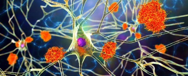
Amyloid plaques in Alzheimer’s disease (Kateryna Kon/Science Photo Library/Getty Images)
A study of around 500,000 medical records has suggested that severe viral infections like encephalitis and pneumonia increase the risk of neurodegenerative diseases like Parkinson’s and Alzheimer’s.
Researchers found 22 connections between viral infections and neurodegenerative conditions in the study of around 450,000 people.
People treated for a type of inflammation of the brain called viral encephalitis were 31 times more likely to develop Alzheimer’s disease. (For every 406 viral encephalitis cases, 24 went on to develop Alzheimer’s disease – around 6 percent.)
Those who were hospitalized with pneumonia after catching the flu seemed to be more susceptible to Alzheimer’s disease, dementia, Parkinson’s disease, and amyotrophic lateral sclerosis (ALS).
Intestinal infections and meningitis (both often caused by a virus), as well as the varicella-zoster virus, which causes shingles, were also implicated in the development of several neurodegenerative diseases.
The impact of viral infections on the brain persisted for up to 15 years in some cases. And there were no instances where exposure to viruses was protective.
Around 80 percent of the viruses implicated in brain diseases were considered ‘neurotrophic’, which means they could cross the blood-brain barrier.
“Strikingly, vaccines are currently available for some of these viruses, including influenza, shingles (varicella-zoster), and pneumonia,” the researchers write.
“Although vaccines do not prevent all cases of illness, they are known to dramatically reduce hospitalization rates. This evidence suggests that vaccination may mitigate some risk of developing neurodegenerative disease.”
Last year, a study of more than 10 million people linked the Epstein-Barr virus with a 32-fold increased risk of multiple sclerosis.
“After reading [this] study, we realized that for years scientists had been searching – one-by-one – for links between an individual neurodegenerative disorder and a specific virus,” said senior author Michael Nalls, a neurogeneticist at the National Institute on Aging in the US.
“That’s when we decided to try a different, more data science-based approach,” he said. “By using medical records, we were able to systematically search for all possible links in one shot.”
First, the researchers analyzed the medical records of around 35,000 Finns with six different types of neurodegenerative diseases and compared this against a group of 310,000 controls who did not have a brain disease.
This analysis yielded 45 links between viral exposure and neurodegenerative diseases, and this was narrowed down to 22 links in a subsequent analysis of 100,000 medical records from the UK Biobank.
While this retrospective observational study cannot demonstrate a causal link, it adds to the pile of research hinting at the role of viruses in Parkinson’s and Alzheimer’s disease.
“Neurodegenerative disorders are a collection of diseases for which there are very few effective treatments and many risk factors,” said co-author Andrew Singleton, a neurogeneticist and Alzheimer’s researcher and the director of the Center for Alzheimer’s and Related Dementias.
“Our results support the idea that viral infections and related inflammation in the nervous system may be common – and possibly avoidable – risk factors for these types of disorders.”
This study was published in Neuron.
mRNA Covid Injections Causing Some Recipients to Experience Drastic Personality Changes: WHY?
Previously healthy and vibrant people have become “docile” and forgetful post-injection, Fuellmich says
Covid jabs are causing people to experience major psychological abnormalities that mirror the effects displayed by people who have had frontal lobotomies, researchers have warned.
According to disturbing new research, large numbers of people who have received the jab are suffering from both physiological and psychological changes that are leaving experts concerned.
Discernreport.com reports: What we DO know is that spike proteins have been found in the brain of at least one vaccine victim who had an autopsy. It seems likely that the one published autopsy of someone who died following the jabs that included a check of the victim’s brain is not an isolated case. Are the spike proteins from the jabs, Covid-19 itself, or both getting into our brains and changing our personalities? A handful of analysts and doctors discussed this potential on a recent interview.
Here’s an article by Ethan Huff over at Natural News that highlights that discussion as well as the potential reasons why some are seeing these dramatic personality changes. It’s noteworthy that after reading the article, I started asking people I know and trust if they’d noticed anything different. ALL of them had. The consensus was that their vaxxed friends and family seemed more docile. This conclusion was derived independently; I talked to each separately and they all had the same basic response without prompting. I’ll be discussing it on today’s episode of The JD Rucker Show.
mRNA Covid Injections Causing Some Recipients to Experience Drastic Personality Changes: WHY?
Last month during an International Crimes Investigative Committee (ICIC) session, attorney Dr. Reiner Fuellmich interviewed Prof. Sucharit Bhakdi, Prof. Dr. Karina Reiss, Dr. Naomi Wolf, and Dr. Peter R. Breggin about the damaging effects of mRNA “vaccination” for covid. One of the topics they discussed is how the shots damage the small capillaries in the brain, override the blood-brain barrier, and cause extensive brain damage that oftentimes results in extreme personality changes.
Some who take the mRNA shots end up experiencing a broken will, which is not exactly a normal side effect of a “vaccine.” What are these things doing to people to change the way their brain functions? That was the subject of the discussion, which you can watch in full either below or at The Exposé.
https://video.icic-net.com/videos/embed/18907e79-a065-4f6e-853e-3c6c2e859a46
During the interview, Wolf unpacked what the post-injection “breaking of people’s will” looks like in real life while Breggin highlighted the disturbing parallels between what the mRNA injections are doing to the brain and the effects of an actual lobotomy.
It quickly becomes clear from their discussion that the covid injection campaign is, in fact, one of the most brutal and savage crimes against humanity that has ever been committed – and all in the name of “public health,” no less. (Related: Some degree of heart damage occurs in every person who gets covid-jabbed with mRNA.)
Previously healthy and vibrant people have become “docile” and forgetful post-injection, Fuellmich says
One of the things Fuellmich and his wife have noticed personally is that servers at local restaurants who were once full of life and very sharp and interactive are no longer their normal selves. Some of them are constantly forgetting things and having to come back to the table while others are now “docile” when they previously had strong personalities.
Wolf explained that she, too, has noticed this. And there are reasons for it that Bhakdi said is caused by the breaking of the blood-brain barrier and insertion of mRNA into brain tissue. And the plan, Bhakdi further revealed in several presentations he has given, is to eventually make all “vaccines” contain mRNA.
“What people do not understand is that all mRNA vaccines are dangerous and are going to threaten life,” Bhakdi told the audience. “It does not matter whether the vaccine encodes for the spike protein, for the measles protein, for the flu – it does not matter. Why? Because the whole danger of the vaccine stems from the ability of the immune system to recognize non-self.”
The damage this causes to the nervous system is something that can be tangibly measured and observed, which is what the discussion highlights. People who got jabbed are no longer themselves, in many cases, expressing unusual emotions that were not previously part of who they were before the injections.
“People, colleagues of mine after they got injected would be much more dualistic in their thinking, much more rigid – and if you know the structure of the brain, that makes sense,” Wolf further explained about her observations.
“If people’s thinking is more rigid and there’s damage to the neural structures, that seems like something worth asking more questions about. I also knew that people were much angrier, less ability to modulate emotions – more primal reactions to provocation. People have also been saying that the changes are affective – people who were previously warm and affectionate have become cold, distant, or cutting other people off.”
https://www.naturalnews.com/2022-12-20-plenty-of-evidence-covid-vaccines-damage-brain.html
by: Arsenio Toledo
Tuesday, December 20, 2022
The evidence overwhelmingly points to the fact that the Wuhan coronavirus (COVID-19) vaccines can cause brain damage.
However, so-called health experts are still denying all evidence supporting the possibility that the COVID-19 vaccines can damage the brain. (Related: New study: mRNA COVID-19 vaccines can cause brain diseases.)
“No, there is no solid evidence to back up this claim,” said Dr. Kelly Cawcutt, an infectious diseases expert at the University of Nebraska Medical Center, referring to a claim that the COVID-19 vaccine can kill brain cells.
“COVID-19 vaccines do not go into your body and acutely kill brain and heart cells. What matters is how a person’s body interacts with the spike protein, either from the infection itself or the vaccine.” She added that cases showing how the vaccine has affected the brain are isolated incidents and do not prove that vaccines affect the brains of the general vaccinated public.
Ben Armstrong of The New American thinks otherwise.
“Can the vaccine damage the brain? There’s been plenty of evidence that it can,” Armstrong said during a recent episode of “The Ben Armstrong Show.” He did acknowledge that COVID-19 vaccine-induced brain damage is not as common as its more popular adverse events such as heart inflammation.
COVID-19 vaccine materials found in the brain
Rhoda Wilson, writing for The Expose, noted that she believes many people who took COVID-19 vaccines have suffered from brain damage.
In an article, Wilson noted that not a lot of studies have regarded how the COVID-19 vaccine affects brain health and function. She blamed this on the eagerness of regulatory authorities like the United Kingdom’s Medicines and Healthcare Products Regulatory Agency and the United States’ Food and Drugs Administration “to license a product about which information appeared to be lacking.”
There’s also evidence proving that the mRNA in Pfizer and Moderna’s COVID-19 vaccines had been found in the brain, along with other major organs like the heart, lung, liver and testicles.
More studies still need to be done, including one that investigates whether the lipid nanoparticles in the COVID-19 vaccines can carry the mRNA across the blood-brain barrier, the semi-permeable barrier that prevents certain substances in the blood from crossing into the protective fluid covering the central nervous system.
“It is vital to know if this happens because if it does, then all bets are off as to what might happen to the brain,” wrote Wilson. If this does happen, she noted that “the neurons, the brain cells, might be marked as foreign by the body’s immune system. And as more booster jabs are given, the problem will get worse.”
Armstrong said the extent of the damage the vaccine can cause is something that the world will only learn through time. What he is worried about is how this will affect children, who are more vulnerable due to their developing immune systems.
“Some experts, advisors and regulators will tell you that the risks are small, but how can they know that?” said Armstrong. “And what is ‘small?’ They told us that the blood clotting problems were small.”
Learn more about how the COVID-19 vaccines damage the body at VaccineDamage.news.
Watch this episode of “The Ben Armstrong Show” on The New American as host Ben Armstrong discusses the damage the COVID-19 vaccines cause to the brain.
https://www.brighteon.com/embed/e33bc5cd-e94a-495a-a489-53be61a8c44e
This video is from the channel The New American on Brighteon.com.
More related stories:
mRNA COVID injections causing some recipients to experience drastic personality changes: WHY?
mRNA spike proteins found in heart, brain of deceased “fully vaccinated” man.
One of the deadliest brain diseases known to man is a “side effect” of COVID jabs.
Sources include: